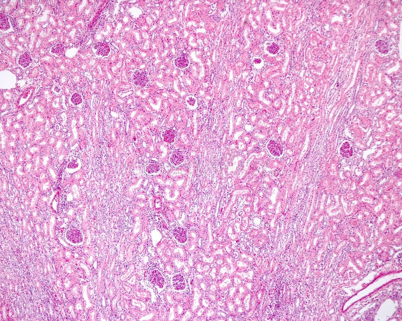
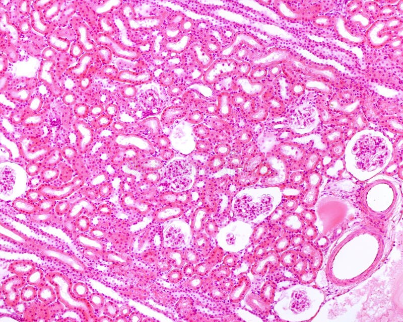
Low magnification micrograph of a renal cortex stained with a silver method to demonstrate reticular fibers. With these techniques, the elements of the nephron are perfectly delimited, since they stain the reticular lamina of the basement membranes. In the image, two medullary rays are identified, formed by tubules parallel and perpendicular to the renal surface. Among them is the cortical
圖片編號:
242706710
拍攝者:
Jlcalvo
點數下載
| 授權類型 | 尺寸 | 像素 | 格式 | 點數 | |
|---|---|---|---|---|---|
| 標準授權 | XS | 480 x 323 | JPG | 13 | |
| 標準授權 | S | 800 x 539 | JPG | 15 | |
| 標準授權 | M | 2110 x 1421 | JPG | 18 | |
| 標準授權 | L | 2724 x 1835 | JPG | 20 | |
| 標準授權 | XL | 3446 x 2321 | JPG | 22 | |
| 標準授權 | MAX | 4561 x 3072 | JPG | 23 | |
| 標準授權 | TIFF | 6450 x 4344 | TIF | 39 | |
| 進階授權 | WEL | 4561 x 3072 | JPG | 88 | |
| 進階授權 | PEL | 4561 x 3072 | JPG | 88 | |
| 進階授權 | UEL | 4561 x 3072 | JPG | 88 |
XS
S
M
L
XL
MAX
TIFF
WEL
PEL
UEL
| 標準授權 | 480 x 323 px | JPG | 13 點 |
| 標準授權 | 800 x 539 px | JPG | 15 點 |
| 標準授權 | 2110 x 1421 px | JPG | 18 點 |
| 標準授權 | 2724 x 1835 px | JPG | 20 點 |
| 標準授權 | 3446 x 2321 px | JPG | 22 點 |
| 標準授權 | 4561 x 3072 px | JPG | 23 點 |
| 標準授權 | 6450 x 4344 px | TIF | 39 點 |
| 進階授權 | 4561 x 3072 px | JPG | 88 點 |
| 進階授權 | 4561 x 3072 px | JPG | 88 點 |
| 進階授權 | 4561 x 3072 px | JPG | 88 點 |


 +886-2-8978-1616
 +886-2-2078-5115


