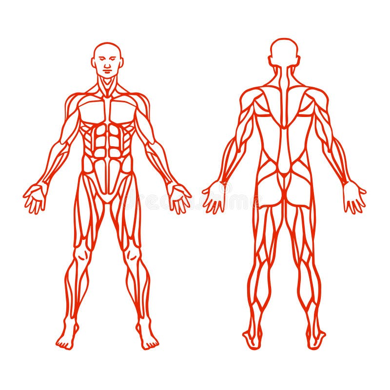
The knee joint is formed by the femur (thigh bone), tibia (shin bone), and patella (kneecap). The ends of these bones are covered in articular cartilage, which provides a smooth surface for the bones to glide against each other during movement.The knee joint is also surrounded by several muscles, which work together to support and stabilize the joint. Some of the key muscles that are involved in knee movement and stability include:Quadriceps: A group of four muscles located in the front of the thigh that work together to extend the knee. These muscles include the rectus femoris, vastus lateralis, vastus intermedius, and vastus medialis.Hamstrings: A group of three muscles located in the back of the thigh that work together to flex the knee and extend the hip. These muscles include the biceps femoris, semitendinosus, and semimembranosus.Gastrocnemius: A muscle located in the back of the calf that works to flex the knee and plantarflex the foot (pushing the foot downward).Popliteus: A small muscle located at the back of the knee joint that helps to unlock the knee joint during the initial stages of knee flexion.
圖片編號:
275691975
拍攝者:
Paihub
點數下載
| 授權類型 | 尺寸 | 像素 | 格式 | 點數 | |
|---|---|---|---|---|---|
| 標準授權 | XS | 480 x 270 | JPG | 13 | |
| 標準授權 | S | 800 x 450 | JPG | 15 | |
| 標準授權 | M | 2309 x 1299 | JPG | 18 | |
| 標準授權 | L | 2981 x 1677 | JPG | 20 | |
| 標準授權 | XL | 3840 x 2160 | JPG | 22 | |
| 標準授權 | MAX | 4618 x 2598 | JPG | 23 | |
| 標準授權 | TIFF | 5431 x 3055 | TIF | 39 | |
| 進階授權 | WEL | 4618 x 2598 | JPG | 88 | |
| 進階授權 | PEL | 4618 x 2598 | JPG | 88 | |
| 進階授權 | UEL | 4618 x 2598 | JPG | 88 |
XS
S
M
L
XL
MAX
TIFF
WEL
PEL
UEL
| 標準授權 | 480 x 270 px | JPG | 13 點 |
| 標準授權 | 800 x 450 px | JPG | 15 點 |
| 標準授權 | 2309 x 1299 px | JPG | 18 點 |
| 標準授權 | 2981 x 1677 px | JPG | 20 點 |
| 標準授權 | 3840 x 2160 px | JPG | 22 點 |
| 標準授權 | 4618 x 2598 px | JPG | 23 點 |
| 標準授權 | 5431 x 3055 px | TIF | 39 點 |
| 進階授權 | 4618 x 2598 px | JPG | 88 點 |
| 進階授權 | 4618 x 2598 px | JPG | 88 點 |
| 進階授權 | 4618 x 2598 px | JPG | 88 點 |






 +886-2-8978-1616
 +886-2-2078-5115


