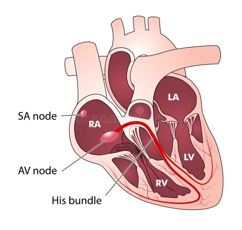
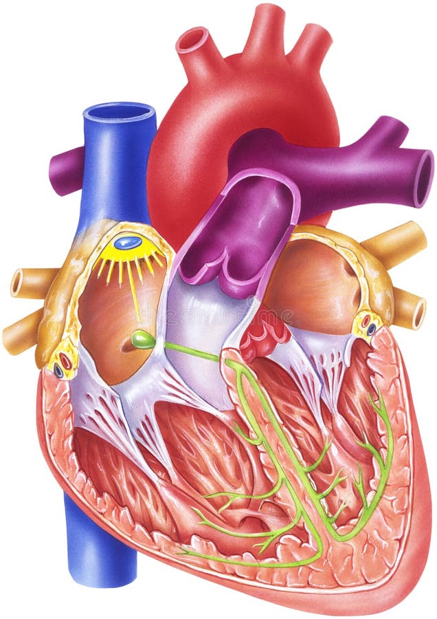
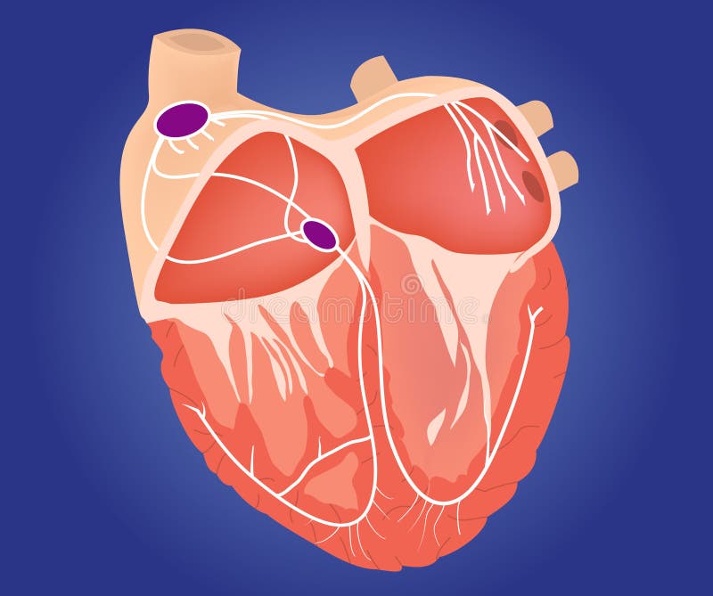
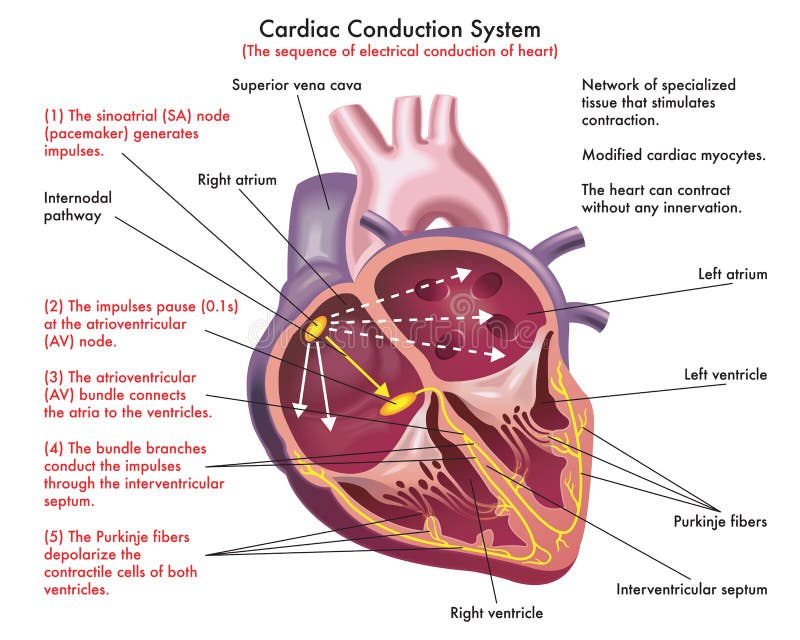
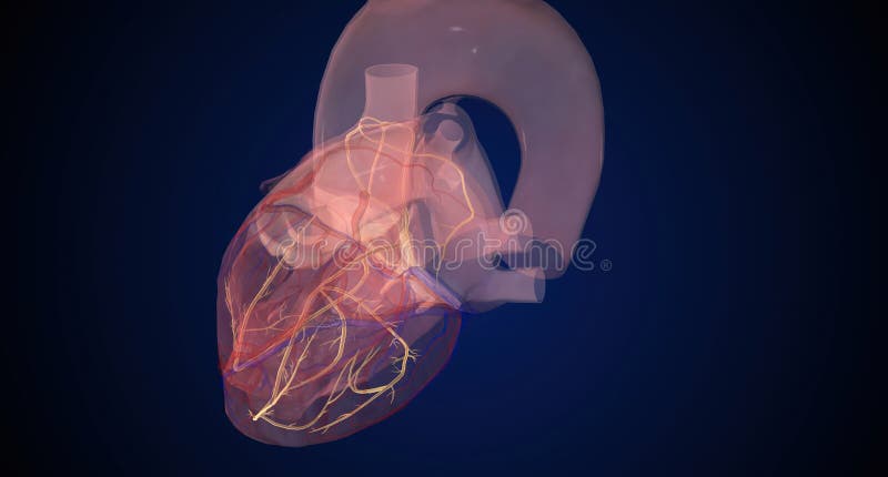
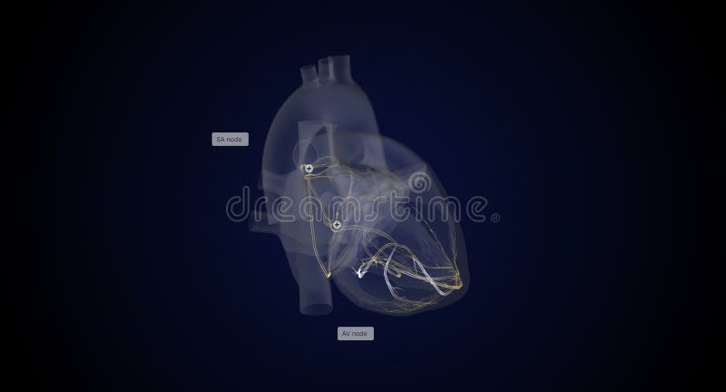
1. The cardiac cycle begins at the SA sinoatrial node located in the sulcus terminalis between the superior vena cava and the right atrium. 2. From this pacemaker, a wave of negativity excitation spreads over both atria and initiates atrial contraction, thereby increasing atrial blood pressure. 3. When atrial pressure exceeds ventricular pressure, both atrioventricular AV valves open and blood rushes into both ventricles. Soon the impulse reaches the AV node and is passed along the AV bundle shown in green to the two ventricles, causing them to contract. 4. When ventricular pressure exceeds atrial pressure, the AV valves close, and this can be heard with a stethoscope as the first of the two heart sounds of the heartbeat. 5. Continued ventricular contraction forces the pulmonary and aortic valves to open, and blood rushes simultaneously into the pulmonary artery and the aorta. 6. When the pressure in these vessels exceeds ventricular pressure, blood tends to rush back into the ventricles, but it gets trapped in the sinuses behind the semilunar cusps. This closes both the pulmonary and aortic valves, resulting in the second of the two heart sounds.
圖片編號:
82100045
拍攝者:
Medicalartinc
點數下載
| 授權類型 | 尺寸 | 像素 | 格式 | 點數 | |
|---|---|---|---|---|---|
| 標準授權 | XS | 339 x 480 | JPG | 13 | |
| 標準授權 | S | 565 x 800 | JPG | 15 | |
| 標準授權 | M | 1455 x 2061 | JPG | 18 | |
| 標準授權 | L | 1879 x 2660 | JPG | 20 | |
| 標準授權 | XL | 2376 x 3365 | JPG | 22 | |
| 標準授權 | MAX | 6456 x 9142 | JPG | 23 | |
| 標準授權 | TIFF | 9130 x 12929 | TIF | 39 | |
| 進階授權 | WEL | 6456 x 9142 | JPG | 88 | |
| 進階授權 | PEL | 6456 x 9142 | JPG | 88 | |
| 進階授權 | UEL | 6456 x 9142 | JPG | 88 |
XS
S
M
L
XL
MAX
TIFF
WEL
PEL
UEL
| 標準授權 | 339 x 480 px | JPG | 13 點 |
| 標準授權 | 565 x 800 px | JPG | 15 點 |
| 標準授權 | 1455 x 2061 px | JPG | 18 點 |
| 標準授權 | 1879 x 2660 px | JPG | 20 點 |
| 標準授權 | 2376 x 3365 px | JPG | 22 點 |
| 標準授權 | 6456 x 9142 px | JPG | 23 點 |
| 標準授權 | 9130 x 12929 px | TIF | 39 點 |
| 進階授權 | 6456 x 9142 px | JPG | 88 點 |
| 進階授權 | 6456 x 9142 px | JPG | 88 點 |
| 進階授權 | 6456 x 9142 px | JPG | 88 點 |









 +886-2-8978-1616
 +886-2-2078-5115


